
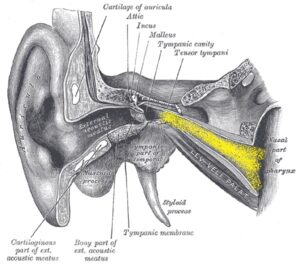
This is just a quick video post of eustachian tube anatomy that we are able to capture this week. There is a little bit of blood in the patient’s nasopharynx here from procedure we did at the same setting just before we made the video. This is the patient’s right eustachian tube, we are passing the scope through the right nasal passage. You can see the large inferior turbinate hanging down into the nasal passage, and just behind the turbinate is the eustachian tube orifice on the left side of the screen. If you look carefully can actually see pulsation of the carotid artery heading up into the central skull base and into the brain. The eustachian tube opening is essentially just in front of this area.
Eustachian tube dilation is most commonly done in the office under local anesthesia. We do have to check a pre-procedure CT scan of the anatomy relative to the middle ear and the carotid artery passageway. We also of course will check a good hearing test to ensure we know what we are dealing with to start with.
Eustachian tube dilation can be helpful to treat chronic eustachian tube pressure, middle ear pressure, problems with plane flights, problems with scuba diving, and ear infections. We have been doing this for a few years in the office, but we finally have some traction with getting insurance companies to cover the cost.