
Thyroid Ultrasound and Biopsy
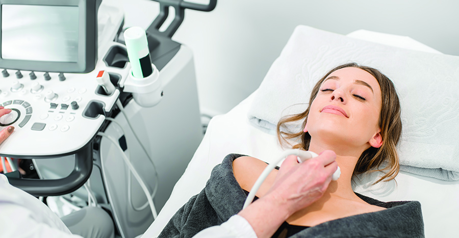
Ultrasound imaging of the thyroid gland is one of the first steps in assessing a thyroid nodule or an enlarged thyroid. Ultrasound is the preferred imaging modality for the thyroid gland, and is performed in the office by your thyroid surgeon. Ultrasound does not use radiation, and is safe for everyone (including babies).
Fine Needle Aspiration Biopsy.
Thyroid nodules are concerning as they can be malignant (cancer). However, approximately 90% of thyroid nodules are completely benign, and require no treatment. The purpose of a fine needle aspiration is to determine the risk of a nodule being malignant, because malignant nodules need to be removed surgically.
If a thyroid nodule is found that has certain size or imaging characteristics, a fine needle aspiration biopsy will be recommended. This procedure involves using an ultrasound to place a fine needle (the diameter of a pin) inside the nodule to obtain a sample of cells. The cells are then examined under a microscope by a pathologist (physician trained in the microscopic appearance of cells), and if necessary, the cells can be processed to determine the presence of genetic mutations for an even more accurate risk analysis. Local (injected) anesthetic is generally all that is necessary, and the procedure takes just a few minutes.
While fine needle aspiration is very useful, the accuracy decreases as the size of the nodule increases. Nodules that are very large (>4 cm) require surgical excision because the risk of a false negative fine needle aspiration biopsy (benign biopsy, when in fact cancer is present) is about 15%.
For more information on thyroid and parathyroid diseases, visit www.thyroidandparathyroid.com.